


Hip dysplasia in dogs doesn’t always announce itself loudly. In many dogs, it develops quietly — slowly changing the way they move, rest, and play before any obvious pain becomes visible. That’s what makes early diagnosis so important, and also so challenging. Professional vets use a step-by-step process that combines physical observation, hands-on examination, and diagnostic imaging to catch the condition before it causes lasting damage.
Here’s a clear breakdown of how that diagnostic process actually works.
1. Breed and Risk Assessment
Every diagnostic process begins with context. Certain breeds carry a much higher genetic risk for hip dysplasia — German Shepherds, Labrador Retrievers, Rottweilers, and Saint Bernards are among the most commonly affected. A vet will ask about the dog’s breed, age, lineage (if known), and any family history of orthopedic issues.
Large-breed puppies are often assessed as early as four months old, especially if the owner reports any hesitation during play, stiffness after rest, or reduced activity. This context helps the vet determine how urgently further screening is needed.
2. Owner-Reported Symptom Review
The owner is often the first to notice something is off. Vets take a detailed history of any behavioral or physical changes the owner has observed. These reported signs often point directly toward hip-related issues.
Being aware of the early signs of hip dysplasia in dogs before the vet visit helps owners provide more accurate histories and catch subtle changes that might otherwise go unmentioned. MedcoVet’s detailed guide is a useful resource for any dog owner wanting to stay informed.
Common owner-reported concerns that prompt hip evaluation:
● Reluctance to jump up, climb stairs, or get into the car
● A ‘bunny hopping’ gait when running
● Stiffness or limping after lying down
● Visible muscle loss in the hindquarters
● Sensitivity when the lower back or hips are touched
These details guide the physical examination and help the vet know exactly where to focus.
3. Physical and Orthopedic Examination
This is where the hands-on assessment begins. The vet will watch the dog walk, trot, and if possible, run — observing stride length, symmetry, and any favoring of one side. They’ll look for the characteristic ‘sway’ or hip drop that often accompanies the condition.
Specific orthopedic tests used during this stage include:
● Ortolani Test — detects hip laxity by feeling for a click or ‘clunk’ as the joint reduces
● Barden Test — measures the degree of lateral movement in the femoral head
● Range of Motion Check — assesses pain or resistance when the hip is extended or flexed
● Palpation — identifies areas of muscle atrophy, joint swelling, or crepitus (grinding sensation)
These tests require sedation in some dogs, particularly those that are tense or in significant discomfort.
4. Radiographic Imaging
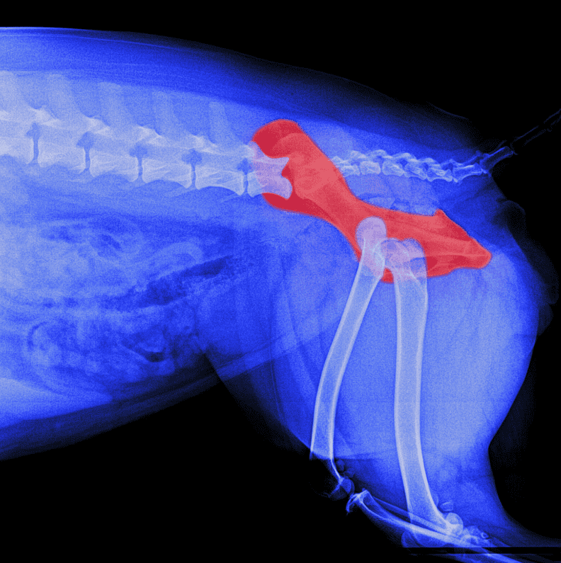
X-rays remain the primary imaging tool for hip dysplasia diagnosis. Standard positioning — called the hip-extended view — lays the dog on their back with legs stretched out, allowing the vet to assess joint space, femoral head shape, and acetabular coverage.
According to the Orthopedic Foundation for Animals (OFA), over 70% of dogs officially evaluated for hip dysplasia show some degree of abnormality. OFA scoring classifies hips as Excellent, Good, Fair, Borderline, Mild, Moderate, or Severe — giving owners and vets a clear framework for next steps.
5. Advanced Imaging When Needed
In some cases, standard X-rays don’t tell the full story. When a dog has inconclusive radiograph results but persistent symptoms, vets may recommend additional imaging.
Advanced options include:
● CT Scans — offer 3D visualization of joint shape and early arthritic changes
● MRI — useful for assessing soft tissue damage around the hip joint
● PennHIP Evaluation — measures distraction index to quantify joint laxity precisely
These tools are typically reserved for cases where surgery is being considered, or when a definitive diagnosis is needed for breeding decisions.
6. Arriving at a Diagnosis and Treatment Plan
Once all the data is collected, the vet puts the picture together. They weigh the physical exam findings, imaging results, symptom history, and the dog’s age and overall health to arrive at a diagnosis and recommend a course of action.
Treatment recommendations vary based on severity:
● Mild cases — weight management, low-impact exercise, joint supplements, anti-inflammatories
● Moderate cases — physiotherapy, hydrotherapy, pain medication management
● Severe cases — surgical options such as TPO, FHO, or total hip replacement
The earlier the diagnosis, the more options are available. Dogs identified at a young age often respond very well to conservative management and can live comfortable, active lives with the right support.
Hip Dysplasia in Dogs – The Conclusion
The diagnostic process is thorough for a reason — hip dysplasia exists on a wide spectrum, and treatment decisions matter. If your dog belongs to a high-risk breed, or if you’ve noticed any subtle changes in how they move or behave, scheduling a veterinary evaluation sooner rather than later is always the right call. Early action gives your dog the best possible chance at a long, pain-free life.