

Have you ever stopped to think about how we all feel or experience certain things in the same way as others?
How do you know the color you perceive as being “red” is the same “red” as the person next to you?
What if their red is your green?
While we can’t answer these mind-boggling questions completely, we can explore the brain’s role in processing external stimuli, like colors, textures, sounds, and so on.
This is where your samatosensory cortex (sometimes referred to as the somatosensory cortex, instead) comes into play.
Responsible for processing external stimuli (or sensations), it plays an integral role in our day-to-day lives.
Below, we will explore this cortex in more detail, including how it works and what role it potentially plays in prosocial behavior.
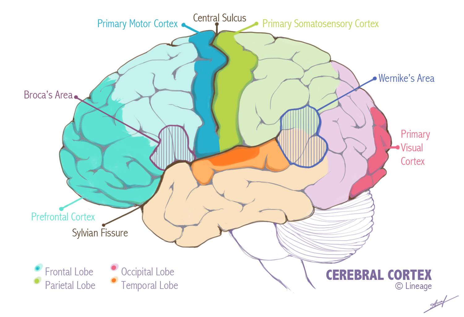
The Location of the Somatosensory Cortex
Before we dive into the important role of the samatosensory cortex, it’s important to understand where it is in your brain and how it contributes toward your brain’s overall anatomy.
It goes without saying that your brain is the central hub of your body. And in order to provide so many different functions, it is a complex structure.
Made up of two sides (or lobes), your brain can be divided into the left- and right-hand side, both of which are connected by the corpus callosum. A different function is performed by each lobe.
The cerebral cortex makes up the outer layer of your brain, acting almost like the skin on a piece of fruit. Its role is to help with processing and more complex thinking skills, like interpreting the environment, language, and reasoning.
Making up part of this cerebral cortex is the somatosensory cortex, which you’ll find in the middle of your brain.
What’s the Role of the Somatosensory Cortex?
The samatosensory cortex receives all of your body’s sensory input. And the cells (or nerves) that extend around your body from the brain are known as neurons.
These neurons sense many different things, including audio, visual, pain, and skin stimuli, and send this information to be processed in the somatosensory cortex. However, the location the neurons send this information to in the cortex isn’t random. Rather, each will have a specific place that’s relevant to the type of information being processed.
When these receptors detect a sensation, they send the information through to the thalamus (the part of your brain that relays receptors’ sensory impulses to the cerebral cortex) before they are passed on to the primary somatosensory cortex.
Once it arrives there, the cortex gets to work interpreting the information. Think of it like any type of data that’s sent to someone for analysis.
Furthermore, some of these neurons are incredibly important, which is why a large portion of this cortex is devoted to understanding and processing all of the information from these neurons. For example, high-level data will be analyzed in more detail and will take more time to interpret, while low-level data will go to a less-equipped analyst, requiring less time to be spent on it.
We can explore this in more detail by using Brodmann’s areas.
Brodmann’s Areas for the Somatosensory Cortex
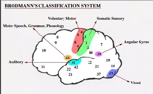
When examining the brain, Korbinian Brodmann, a German neurologist, identified 52 different regions according to how different their cellular composition was. Today, many leading scientists will still use these areas, hence why they are often referred to as “Brodmann’s areas.”
When it came to the somatosensory cortex, Brodmann divided this into four areas, 1, 2, and 3 (which is further divided into 3a and 3b).
These numbers were assigned by Brodmann based on the order he examined the area, and, therefore, are not indicative of their importance.
After all, area 3 is often seen as the primary area of this cortex.
How come?
Area 3 is responsible for receiving the bulk of the input that comes straight from the thalamus, with the information being processed initially in this area.
Area 3b is concerned specifically with the basic processing of things we touch, while 3a responds to the information that comes from our proprioceptors (these are specialized sensors that are located on the ends of your nerves that are found in joints, tendons, muscles, and the inner ear, relaying information about position or motion so you are constantly aware of how your body is moving or is positioned in a space).
Areas 1 and 2 are densely connected to 3b.
Therefore, while the primary location for any information about the things we touch is sent to 3b, it will also be sent to areas 1 and 2 for further in-depth processing.
For example, area 1 appears to be integral to how we sense the texture of something, while area 2 seems to have a role in how we perceive this object’s shape and size. Area 2 also plays a role in proprioception (this enables us to orientate our bodies in a particular environment without us having to consciously focus on where we are).
Should there be any lesions to these areas of the cortex (those that support the roles mentioned above, in particular) then we may notice some deficits in our senses. For example, if there is a lesion to area 1, we will find a shortfall in our ability to distinguish the texture of things, while a lesion to area 3b will affect our tactile sensations.
Somatotopic Arrangement
Each of the four areas we have mentioned are arranged in such a way that a particular area will receive information from a specific part of the body. This is what is known as the somatotopic arrangement, with the entire body being represented within each of the four areas of the somatosensory cortex.
And as some parts of our bodies are more sensitive, e.g. the hands and lips, this requires more cortex and circuitry to be dedicated to processing any sensations that come from these areas. Therefore, if you look at somatotopic maps that depict the somatosensory cortex, you will notice they are distorted, with the areas of the body that are highly sensitive taking up far more space in this area.
How the Samatosensory Cortex May Contribute in Prosocial Behavior
As we now know, when someone experiences pain, this bodily sensation is processed in their brain. It will also switch on an emotional reaction in their brain, too.
However, when we see someone else in this type of pain, many of these same regions are activated in our own brains. But this differs entirely when you are dealing with a convicted criminal with psychopathic tendencies.
When they see someone else in pain, there is less activation in these specific areas of the brain. They will also show disregard and less empathy toward others.
What does this suggest?
That when these “shared activations” are lacking it can cause issues with a person’s empathy.
In fact, over the years, scientists have developed the belief that we are able to feel empathy for others who are in pain because of these shared activations – and this is why we have a desire to help them.
That said, there is still a lack of evidence which helps identify how helpful behavior is influenced by these pain-processing areas of our brain. That’s why some suggest that helpful behavior is contributed to very little by empathy-related processes.
Further Studies
To explore this further, one study looked at participants’ reactions to a video of someone being swatted on their hand by a belt while displaying different levels of pain. The participants could then indicate how much pain they felt this person was in by donating money to them – so the more pain they thought they were in, the more money they donated to try and ease this.
Throughout the study, the participants’ brains (their samatosensory cortex, in particular) were measured. And the results found that the more activated this area was, the more money they donated.
The researchers then interfered with the participants’ brain activity using various techniques that affected how they perceived the sensations in their hand. This altered their accuracy in assessing the pain of the victim, and it also caused disruption to the link between the perceived pain of the victim and the donations. The amount of money being given was no longer correlating to the pain they were witnessing.
A Role in Social Function
These findings suggest that the area of the brain that helps us perceive pain (the somatosensory cortex) plays a role in our social function. It helps us transform the vision of bodily harm into an accurate perception of how much pain the other person is experiencing. And we need these feelings in order to adapt so we can help others.
This also adds to the current argument of what role empathy plays in helping behaviors, with it suggesting that we are indeed promoted to help by brain activity that is empathy-related. It allows us to pinpoint who needs our help.
Putting These Findings into Practice

By understanding this relationship between the activity in our brain and our helping behavior, it may help in the development of treatments for people who are suffering with antisocial behavior. Or for children with unemotional, callous traits – something that’s associated with a general disregard for other people and a lack of empathy.